
A patient-advocate companion guide to the graphic of the same title.
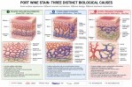
Same surface appearance. Different vascular architecture. Different biology. Different treatment implications.
[ FULL GRAPHIC ]
What “different biology underneath” looks like — the same surface, three different vascular architectures —
Three [of Many] Distinct Causes of a Port Wine Stain ◈ →
Before You Read
I was born with a port wine stain covering both legs and most of my trunk — from hip to toe.
For more than forty years, doctors called it a hemangioma, a flamed nevus, and a port wine stain. None of those labels led to the right treatment, because none of them led to a proper diagnosis. Doctors prescribed heavy compression garments. Those garments caused lifetime injury and permanent disabilities — because no one had confirmed what was actually underneath the skin before deciding what to do about what was on top of it.
I was finally diagnosed with Klippel-Trenaunay Syndrome at age 42, by a skilled vascular surgeon who was weeks from retirement. He looked at the imaging. He saw what the labels had missed. Among his findings: no deep veins. Compression garments were not just unhelpful. They were contraindicated.
I tell you this not to frighten you. I tell you this because it is the reason this graphic exists — and the reason this guide accompanies it.
A port wine stain is not a diagnosis. It is a visible clue.
Why the Same Birthmark Can Mean Different Things
Port wine stains are congenital vascular anomalies present from birth. They do not fade on their own and tend to darken over time. That much is shared across all presentations.
What is not shared is what is happening underneath the skin.
The same reddish-purple surface appearance can result from at least three distinct biological causes — and those differences matter enormously for treatment. The graphic this guide accompanies was created to make that point visible. What follows explains what it shows.
The Three Causes
1 — Isolated Capillary Malformation Classic Port Wine Stain — Slow Flow
The most common presentation. Mildly dilated capillaries in the dermis, normal venous structure, no arteriovenous shunting, lymphatics typically unaffected. The architecture is relatively straightforward. Most isolated port wine stains involve a somatic mutation in the GNAQ gene — occurring in a single cell during early development, not inherited from a parent.
Pulsed-dye laser therapy has its strongest evidence base in this population. Even here, complete clearance is rare. Lightening is the realistic goal.
Syndromic associations are uncommon — but a birthmark involving the forehead and upper eyelid raises the possibility of Sturge-Weber Syndrome and warrants specialist evaluation before any assumptions are made.
Before treatment: Is this truly isolated? Has deeper venous, lymphatic, or neurological involvement been ruled out by imaging?
2 — Sturge-Weber Syndrome Capillary–Venous Malformation — Slow Flow
Here the port wine stain is the surface expression of something happening in the brain and eyes as well. Abnormal blood vessel networks develop on the brain surface and in the eye, driven by the same GNAQ mutation — occurring randomly during embryonic development, not inherited.
Under the skin, the port wine stain in Sturge-Weber is driven by venous pressure: engorged, dilated vessels under elevated pressure from impaired drainage below. This is the vascular architecture depicted in the widely shared educational illustration that originally prompted this body of work.
Brain and eye involvement require their own monitoring — neurology for seizures, ophthalmology for glaucoma. Laser therapy has a theoretical fit with the Sturge-Weber pathology. Clinical outcomes are more sobering than the theory suggests.
Before treatment: Has Sturge-Weber been ruled in or out with MRI and ophthalmologic evaluation? Has the laser recommendation been calibrated to Sturge-Weber outcomes specifically?
3 — Klippel-Trenaunay Syndrome Capillary–Venous–Lymphatic Malformation — Slow Flow
In KTS, the port wine stain is the most consistent and visible feature — present in approaching 100% of patients — but it reflects a different biological reality than either of the above. It is not driven by engorged venular pressure. It reflects a capillary differentiation failure, driven by overactivation of the PI3K/mTOR pathway, most commonly from a somatic PIK3CA mutation.
The venous and lymphatic components beneath the skin vary enormously across the KTS spectrum — from severe to mild to barely detectable. Many KTS patients, particularly those with bilateral limb and trunk involvement, do not have the pressurized venular architecture that laser therapy is designed to target.
In PIK3CA-driven KTS, the genetic program that builds abnormal vessels does not switch off after laser treatment. Surface lightening may occur. The underlying instruction does not change. Recurrence is biologically expected, not a treatment failure.
Before treatment: Has imaging confirmed what is actually beneath the skin? Has the genetic profile been considered? What are realistic outcome expectations for this specific diagnosis — not for port wine stains generally?
What This Graphic Is Asking You to Do
Look at the three panels side by side. Same skin color. Three different structures underneath.
Then ask one question before any treatment decision is made:
Which one of these is my child?
Modern medicine has the tools to answer that question. Doppler ultrasound. MRI. Genetic testing. Specialized vascular anomaly centers. These tools exist precisely because the surface appearance is not enough information to treat from.
A visual diagnosis is a starting point. Imaging is the next step. A specialist in vascular anomalies — ideally at an ISSVA-affiliated center — is the right person to interpret what imaging finds.
Bring this guide and the graphic to that appointment. Ask which biological cause best describes your situation. Then, and only then, make treatment decisions.
What Comes Next
If you have moved past the birthmark labels and started hearing syndrome names — Klippel-Trenaunay, Parkes Weber, Sturge-Weber, KTWS — the next companion guide addresses those directly.
Three conditions that were once grouped under a single diagnosis. Why they were separated. What the difference means for your family.
[ Next: Three Confounded Syndromes → ]
And if you are ready for the full map — every condition the science has named within this landscape, organized and linked — the Vascular Anomalies Compendium is there when you are.
[ Vascular Anomalies Compendium → ]
Care4-Rare is a non-commercial patient advocacy project. Nothing here substitutes for evaluation by a qualified specialist in vascular anomalies. Bring these materials to your doctor — they are designed to support that conversation, not replace it.
Sources
[CM-001] Hammill AM, et al. Capillary Malformations. J Clin Invest. 2024. https://pubmed.ncbi.nlm.nih.gov/38618955/
[SWS-001] NIH/NCBI StatPearls. Sturge-Weber Syndrome. Updated 2026. https://www.ncbi.nlm.nih.gov/books/NBK459163/
[KTS-001] NIH MedlinePlus Genetics. Klippel-Trenaunay Syndrome. https://medlineplus.gov/genetics/condition/klippel-trenaunay-syndrome/
[KTS-002] Alazawi S, Wright K. KTS With Atypical Presentation. Cureus. 2022. https://pmc.ncbi.nlm.nih.gov/articles/PMC9497453/
[GEN-001] Liu L, et al. Pathogenesis of Port-Wine Stains. PMC9603382. 2022. https://pmc.ncbi.nlm.nih.gov/articles/PMC9603382/
See companion: Three Confounded Syndromes | Vascular Anomalies Compendium
