
“They look like us.”
Part 2 of 8 in the Care4-Rare guided introduction.
Preview: The next post shows exactly our different biology which is underneath what we look like — the same surface, three different vascular architectures.
In the sidebar to the right is a a ready, a website graphic. We fully discuss the content in this graphic in the next post
It is one of the most natural things a parent does in the first days after a diagnosis.
You go online. You find a support group, a Facebook page, a forum full of families navigating the same unfamiliar territory. You scroll through photographs. And there — in another child’s face, another child’s leg, another child’s arm — you see something that looks like your child.
The relief that follows is real. The community that follows is real. The sense that someone else understands is real, and valuable, and worth protecting.
What I am asking you to hold alongside that relief is one careful question:
Does looking the same mean being the same?
In most of life, the answer is close enough to yes. In congenital vascular anomalies, modern medicine has learned that the answer is often, crucially, no.
The Same Surface. Different Stories Underneath.
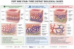
A reddish-purple mark on the skin — what clinicians call a port wine stain, or a capillary malformation — can arise from at least three entirely different biological causes. The same color, the same texture, the same appearance in a photograph. Three different vascular architectures underneath. Three different biological realities, each with different implications for treatment and outcome.
The graphic that follows this post was created to make that visible. It shows all three side by side — what each looks like on the surface, and what is actually happening in the vessels beneath. It is the most important single image on this website for a family encountering a port wine stain for the first time.
What the graphic shows — and what the companion guide explains in full — is this:
A port wine stain is a visible clue. It is not, by itself, a diagnosis.
Modern medicine has the tools to look beneath the surface. Ultrasound. Doppler studies. MRI. Genetic testing. These tools exist specifically because clinicians learned that what they could see was not telling them enough. A doctor looking only at the skin is working with incomplete information. A parent who knows to ask what is underneath is already ahead of where I was at forty-two.
What the Graphic Will Show You
Three panels. Same skin color across all three. Underneath:
One has mildly dilated capillaries and a relatively simple architecture — the most common presentation, and the one for which most laser therapy evidence was developed.
One has engorged vessels under venous pressure, driven by impaired drainage deeper in the tissue — the presentation associated with Sturge-Weber Syndrome, where the brain and eyes are also involved.
One has a capillary malformation overlying a combined venous and lymphatic architecture of varying depth and severity — the presentation associated with Klippel-Trenaunay Syndrome, where the genetic instruction driving abnormal vessel formation continues to operate beneath whatever treatment addresses the surface.
They can look identical on the outside. They are not identical underneath. The companion guide explains the biology, the treatment implications, and the questions worth bringing to a specialist before any decision is made.
Care4-Rare is a non-commercial patient advocacy project. Nothing here substitutes for evaluation by a qualified specialist in vascular anomalies.
Next: Post 3 — “Three of Many Causes” — Our designed graphic comparing three of many causes of Port Wine Stains →
footer
