
We are not the same.
Part 4 of 8 — a visual companion to Part 2: They Look Like Us.
This graphic lives in the sidebar as well — a permanent quick reference for your specialist appointments and your own research.
[ Thumbnail image of graphic — links to full graphic page ]
[ Continue: We Are Not The Same → ]
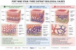
What “different biology underneath” looks like — the same surface, three different vascular architectures —
Three Distinct Causes of a Port Wine Stain ◈ →
It is also in the sidebar whenever you need it.
It is one of the most natural things a parent does in the first days after a diagnosis.
You go online. You find a support group, a Facebook page, a forum full of families navigating the same unfamiliar territory. You scroll through photographs. And there — in another child’s face, another child’s leg, another child’s arm — you see something that looks like your child.
The relief that follows is real. The community that follows is real. The sense that someone else understands is real, and valuable, and worth protecting.
What I am asking you to hold alongside that relief is one careful question:
Does looking the same mean being the same?
In most of life, the answer is close enough to yes. In congenital vascular anomalies, modern medicine has learned that the answer is often, crucially, no.
The same surface. Different stories underneath.
A reddish-purple mark on the skin — what clinicians call a port wine stain, or a capillary malformation — can arise from at least three entirely different biological causes.
In the most common presentation, it is an isolated capillary malformation: dilated blood vessels close to the skin’s surface, without significant involvement of deeper structures. The visible mark is essentially the whole story.
In Sturge-Weber Syndrome, that same surface appearance is the visible sign of something happening in the brain and eyes as well — abnormal blood vessel development driven by a specific genetic mutation, with implications for seizures, glaucoma, and neurological development that have nothing to do with the color on the skin.
In Klippel-Trenaunay Syndrome, the capillary mark may be accompanied by venous and lymphatic malformations, and overgrowth of soft tissue or bone — a combined condition that behaves very differently from either of the above, with different risks, different imaging findings, and different appropriate care.
These three can look identical on the surface. Beneath the surface, they are different diseases.
This is what the first graphic shows.
The illustration titled “Port Wine Stain: Three Distinct Biological Causes” was created to make this visible. Same surface color. Three different vascular architectures underneath. Three different biological realities.
Look at it not as a diagram of disease but as a diagram of why two children who appear to have the same thing may need very different care — and why the visual similarity that first brought you comfort in a support group is the beginning of the conversation, not the end of it.
A doctor looking only at the skin is working with incomplete information. A parent who knows to ask what is underneath is already ahead of where I was at forty-two.
A port wine stain is a visible clue. It is not, by itself, a diagnosis.
The good news embedded in this complexity is real: modern medicine has the tools to look beneath the surface. Ultrasound. Doppler studies. MRI. Genetic testing. Specialized vascular anomaly centers. These tools exist specifically because clinicians learned that what they could see was not telling them enough.
The next post takes this one step further — into the history of how the syndrome labels themselves have shifted, split, and been refined over time, and what that means for families navigating a diagnosis today.
Next: Post 3 — “We are not the same” — how one syndrome became three →
footer
We are not the same.
Part 3 of 8 — a visual companion to Part 2: They Look Like Us. [ ← They Look Like Us ] [ We Are Not The Same → ]
This graphic lives in the sidebar as well — a permanent quick reference for your specialist appointments and your own research.
[ Thumbnail image of graphic — links to full graphic page ]
[ Continue: We Are Not The Same → ]
For most of the twentieth century, a patient who presented with a port wine stain, an enlarged limb, and abnormal veins received a single diagnosis: Klippel-Trenaunay-Weber Syndrome.
The physicians who made that diagnosis were not careless. They were working with the tools available to them — principally their eyes, their hands, and their clinical judgment. What they could observe was a pattern. A recurring constellation of visible features. And they named it.
What they could not always observe was what the blood vessels were actually doing underneath.
They were classifying what they could see. Modern medicine increasingly classifies what the vessels are actually doing.
— — —
The tool that changed everything.
When Doppler ultrasound became widely available — and later MRI, MR angiography, and detailed flow studies — physicians were able, for the first time, to characterize not just the anatomy of abnormal vessels but their physiology. Their behavior. Their flow.
And what they found was that the patients who had all been grouped under a single label were not, in fact, the same.
Some patients had slow-flow malformations: dilated capillaries, abnormal veins, lymphatic involvement. Blood moving sluggishly through vessels that were malformed but not high-pressure. This is what we now recognize as Klippel-Trenaunay Syndrome.
Other patients had something fundamentally different: arteriovenous shunting. Arterial blood — blood under high pressure, designed to travel through arteries — bypassing the normal capillary bed and entering the venous system directly. This is high-flow physiology. It carries different risks, produces different symptoms, and requires different treatment entirely. This is what we now recognize as Parkes Weber Syndrome.
Same skin. Same enlarged limb. Same abnormal veins visible on the surface. Completely different vascular biology underneath.
— — —
Why this matters for your family today.
The second graphic — “Three Syndromes on the Klippel-Trenaunay Spectrum” — exists to make this visible. It shows KTS, Parkes Weber with arteriovenous malformation, and Parkes Weber with arteriovenous fistula side by side: what each looks like on the surface, and what the vascular architecture beneath each diagnosis actually is.
The reason this history matters is not academic. It is practical.
If a family receives a diagnosis of Klippel-Trenaunay Syndrome today — and some clinicians still use the older combined label Klippel-Trenaunay-Weber Syndrome — the question worth asking is: has the flow been characterized? Has imaging confirmed whether this is slow-flow or high-flow? Has arteriovenous shunting been ruled in or ruled out?
Because therapies appropriate for slow-flow venous and lymphatic malformations can be ineffective — or harmful — when applied to high-flow arteriovenous lesions. The label is not enough. The biology underneath the label is what determines the appropriate care.
The same syndrome label once described several fundamentally different vascular disorders. Knowing which one matters.
We are not the same. Port wine stains are not the same. The syndromes behind them are not the same. The treatments are not the same.
The next post brings this to its natural destination: what a specialist in vascular anomalies is actually thinking when they look at your child — and why the studies they order are not excessive caution but necessary science.
Next: Post 4 — What the specialists are actually looking at →
